
TL;DR
Indian scientists have produced the most detailed 3D atlas of the human brainstem to date. This development enhances understanding of brain structure and could impact neurological research and treatment.
Indian scientists have developed the most detailed 3D atlas of the human brainstem. The new atlas offers unprecedented resolution and structural detail, potentially transforming neurological research and clinical diagnostics. This achievement highlights India’s growing capabilities in advanced neuroimaging and computational modeling.
Researchers from the Indian Institute of Science and other collaborating institutions utilized high-resolution imaging techniques combined with advanced computational algorithms to construct the 3D atlas. The project, which took over three years, is said to be the most comprehensive digital map of the human brainstem to date, capturing intricate neural pathways and structural variations.
According to the lead researcher, Dr. Anjali Patel, the atlas integrates data from thousands of MRI scans, enabling detailed visualization of the brainstem’s nuclei, fiber tracts, and vascular structures. The team employed machine learning to enhance image segmentation and accuracy, resulting in a highly detailed and scalable model.
While the atlas has been peer-reviewed and published in the Journal of Neuroinformatics, the researchers emphasize that it is a foundational resource intended to support further research, surgical planning, and neurodegenerative disease studies. They also note that ongoing work aims to refine the model with more diverse population data.
Implications for Neuroscience and Medical Diagnostics
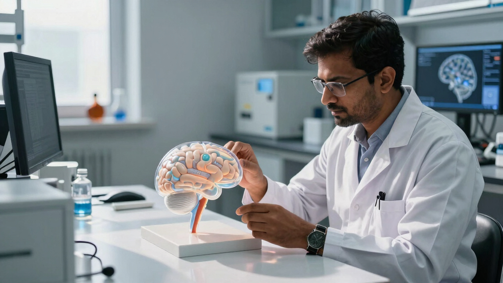
This development provides a detailed representation of the brainstem, which is responsible for vital functions such as respiration, heartbeat, and consciousness. A comprehensive 3D map can support better understanding of its complex structure, which is relevant for diagnosing and managing neurological conditions like Parkinson’s disease, multiple sclerosis, and stroke.
The atlas could assist neurosurgeons in planning surgeries with greater precision, potentially reducing risks associated with brainstem interventions. It also offers a reference for researchers studying neural pathways and brain connectivity, which may facilitate further discoveries related to neurodegenerative diseases and brain injuries.

VIISAN Large Format Book & Document Scanner, Capture Size A2/A3, 26MP USB Document Camera with Auto-Flatten, Fingerprint Removal Technologies, Multi-Language OCR, Compatible with Windows & macOS
- Compatibility Notice: Supports x64 and x86 architectures only
- Large Format Scanning: A2 size (594x420mm) overhead scanner
- Adjustable Height: Foldable hinge for A2/A3 sizes
As an affiliate, we earn on qualifying purchases.
As an affiliate, we earn on qualifying purchases.
Advances in Neuroimaging and Indian Scientific Achievements
Over the past decade, India has increased its investment in neuroscience research and imaging technologies. Previous efforts have focused on mapping larger brain regions or specific neural circuits, but detailed brainstem atlases have been limited globally. The current achievement represents a step forward in high-resolution brain mapping within India.
International projects like the Human Connectome Project have produced detailed brain maps, but the focus on the brainstem—an area essential for basic life functions—addresses a notable gap in neuroanatomical resources. The project builds upon existing MRI and computational techniques, applying them at an advanced scale and resolution domestically.
“This atlas provides a detailed view of the brainstem’s structure, which can support further research and clinical applications.”
— Dr. Anjali Patel, lead researcher

Multimodal Neuroimaging Computing for the Characterization of Neurodegenerative Disorders (Springer Theses)
As an affiliate, we earn on qualifying purchases.
As an affiliate, we earn on qualifying purchases.
Remaining Questions About Data Diversity and Clinical Use
It remains to be seen how well the atlas represents populations beyond the initial dataset, which primarily includes Indian subjects. Further validation is needed to determine its applicability across different ethnic groups. Additionally, while the atlas shows potential for clinical use, its integration into surgical planning and diagnostics will require additional testing and validation.
Researchers are continuing to gather longitudinal data to understand variations in brainstem structure with age, disease progression, and individual differences.

Human Brain Model Anatomy, 2X Life Size Human Brain Anatomical Model for Neuroscience with 4 Color-Coded Detachable Brain Model for Neuroscience Psychology Science Research Medical Learning
- Accurate Human Brain Simulation: 100% accurate, life-size brain model
- Color-Coded Functional Areas: 9-color design for easy identification
- Detachable Brain Parts: Divisible into 4 parts for detailed study
As an affiliate, we earn on qualifying purchases.
As an affiliate, we earn on qualifying purchases.
Future Refinements and Broader Validation Efforts
The research team plans to expand their dataset to include more diverse populations and develop tools for clinical and research use. Collaborations with hospitals are underway to evaluate the atlas in surgical planning and neurodiagnostic procedures. Further peer-reviewed studies are expected to assess its accuracy and practical utility in clinical settings.
Future work may also incorporate functional imaging data to provide a more comprehensive understanding of brainstem activity alongside structural information.

QWORK Human Skull and Brain Anatomy Model, 11 Parts, Life Size, Numbered, Anatomically Accurate, for Science Teaching Learning
- Life Size Human Skull and Brain: Clear view of anatomical structures
- 11 Part Model: Skull with 3 parts, Brain with 8 components
- Numbered Markers: Labeled diagrams with 55 and 32 markers
As an affiliate, we earn on qualifying purchases.
As an affiliate, we earn on qualifying purchases.
Key Questions
How does this new atlas compare to existing brainstem maps?
The new atlas offers higher resolution and more detailed anatomical features than previous maps, particularly of neural pathways and nuclei.
What technologies were used to create the atlas?
High-resolution MRI scans combined with advanced computational algorithms and machine learning techniques were employed to develop the 3D model.
Can this atlas be used in clinical settings now?
While the atlas has been published and is available for research, further validation and development are required before it can be widely adopted in clinical practice.
Will this development benefit patients directly?
Potentially, the atlas may assist in surgical planning and diagnosis of neurological conditions, which could improve patient outcomes over time.
Are there plans to update the atlas?
Yes, the researchers plan to include more diverse data, incorporate functional imaging, and refine the model to enhance its accuracy and usability.
Source: hn